M7 Wifi Camera Notes
I picked up a cheap Wifi security camera on Amazon, it uses a mobile app called “seeing”. Which appears to be that referenced at seeing.store.
No ports were open when scanned with nmap. Monitoring traffic on the router indicated it was talking to a remote server (appears to be on Alibaba’s cloud service):
traffic when streaming video:
18:19:21.995059 IP 192.168.8.191.36198 > 47.79.82.123.443: Flags [F.], seq 228080, ack 6579, win 6169, length 0
18:19:21.998765 IP 47.79.82.123.443 > 192.168.8.191.36198: Flags [F.], seq 6579, ack 228080, win 831, length 0
18:19:22.671581 IP 192.168.8.191.36200 > 47.79.82.123.443: Flags [S], seq 4002814628, win 29200, options [mss 1460,sackOK,TS val 4294938745 ecr 0,nop,wscale 3], length 0
With some more fiddling it appears that the cloud service coordinates with the phone/camera but if they are on the same network the camera will stream directly to the phone:
8:52:29.753976 IP navas-iPhone.lan.49936 > 192.168.8.191.10666: UDP, length 48
...
18:52:29.841608 IP 192.168.8.191.10666 > navas-iPhone.lan.49936: UDP, length 1332
18:52:29.841943 IP 192.168.8.191.10666 > navas-iPhone.lan.49936: UDP, length 1332
Whatever communication the phone coordinates via the cloud appears to open up a UDP port on the camera. It first sends a packet with a payload of “dummy”:

Which the camera seems to echo back:

The phone then sends a more complex binary payload:

The camera then replies with some clear text:

The phone sends some more binary data to setup the stream… I was able to replay the initial packet from a PC and receive the clear text reply. But this only works if the port has been opened via the phone first.
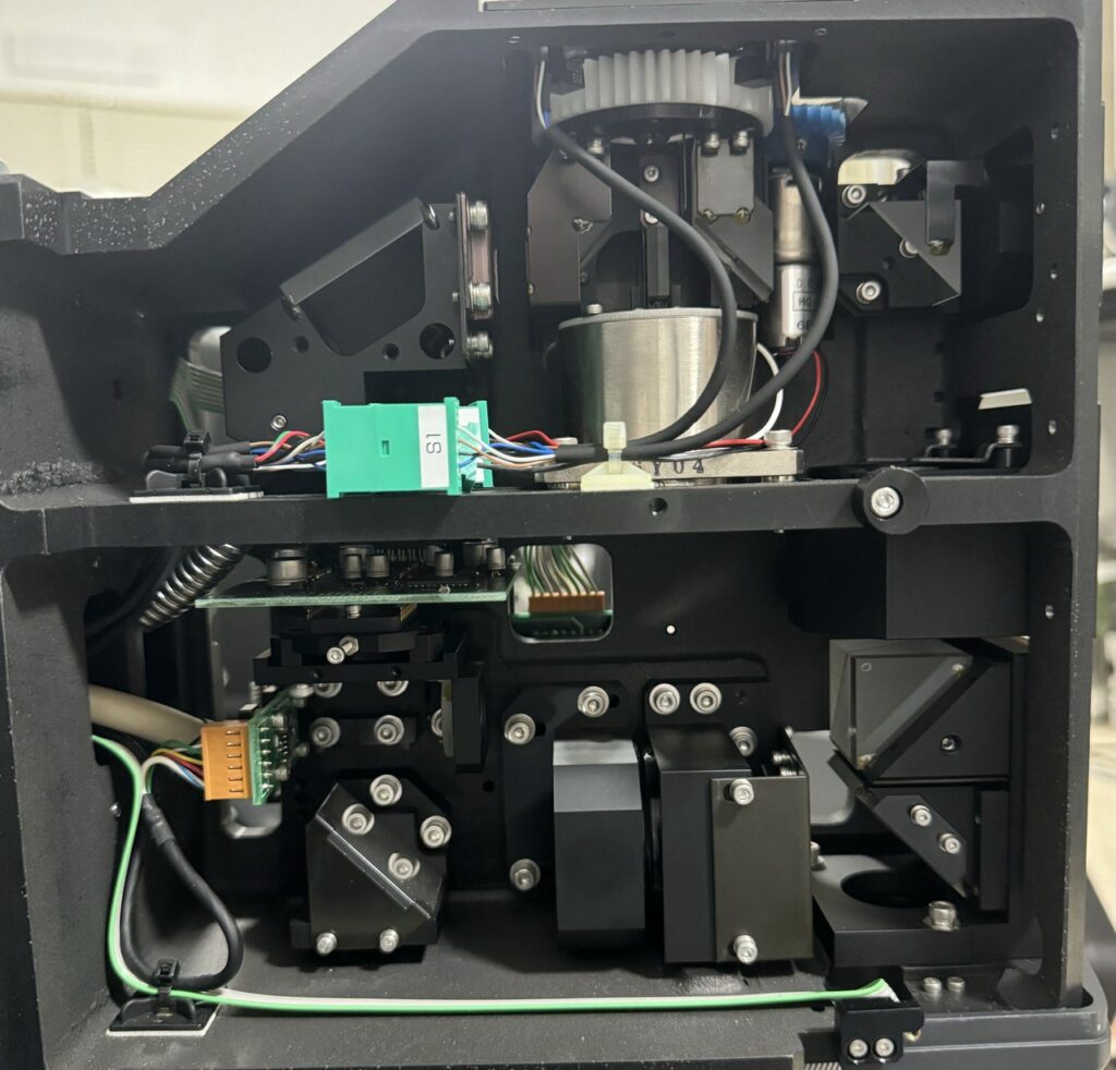
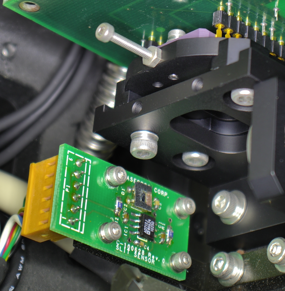
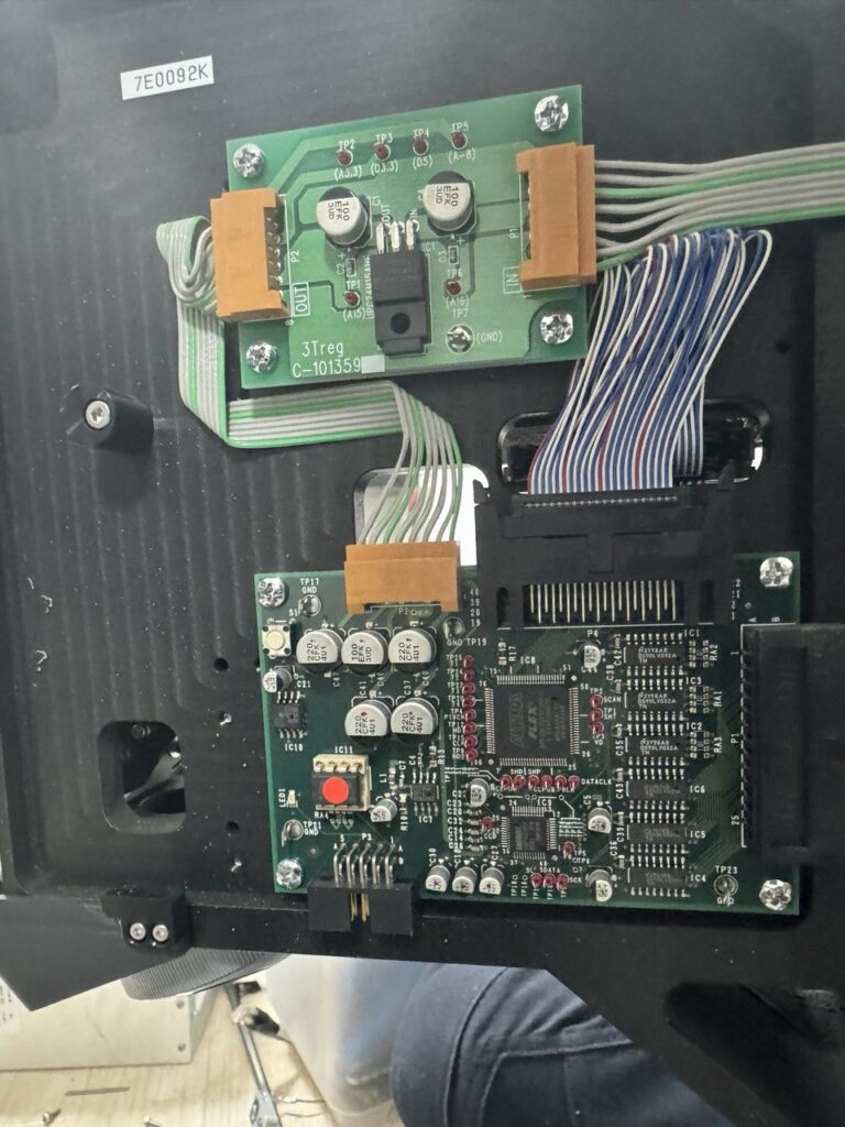
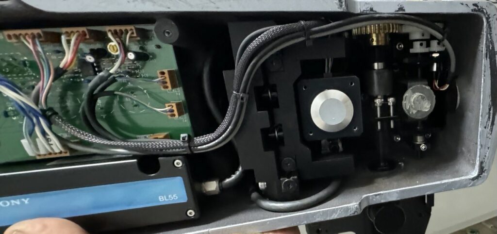
With no clear routes into the camera via the network I pulled it apart. The camera uses a AllWinner IP Camera SoC. I tried probing all the pads labelled RX/TX for a serial interface but didn’t have any luck here.
So, dumped the flash next.
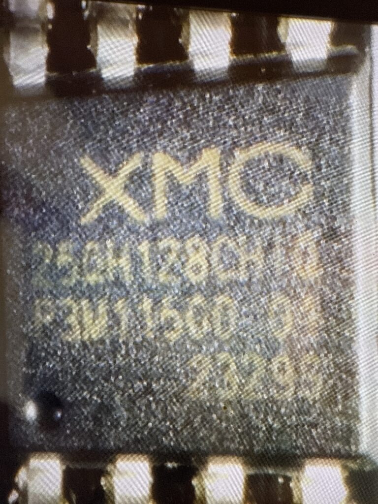
Via a raspberry pi using: sudo flashrom -p linux_spi:dev=/dev/spidev0.0,spispeed=500K -r m7_firmware.bin
The camera appears to use an OpenWRT fork called Tina Linux. eGON header found at beginning of flash, indicating use of Allwinner’s built-in BROM and SPL bootloader. Bootargs suggest a serial console should be present.. so some more hunting may reveal it.
That’s all for now!
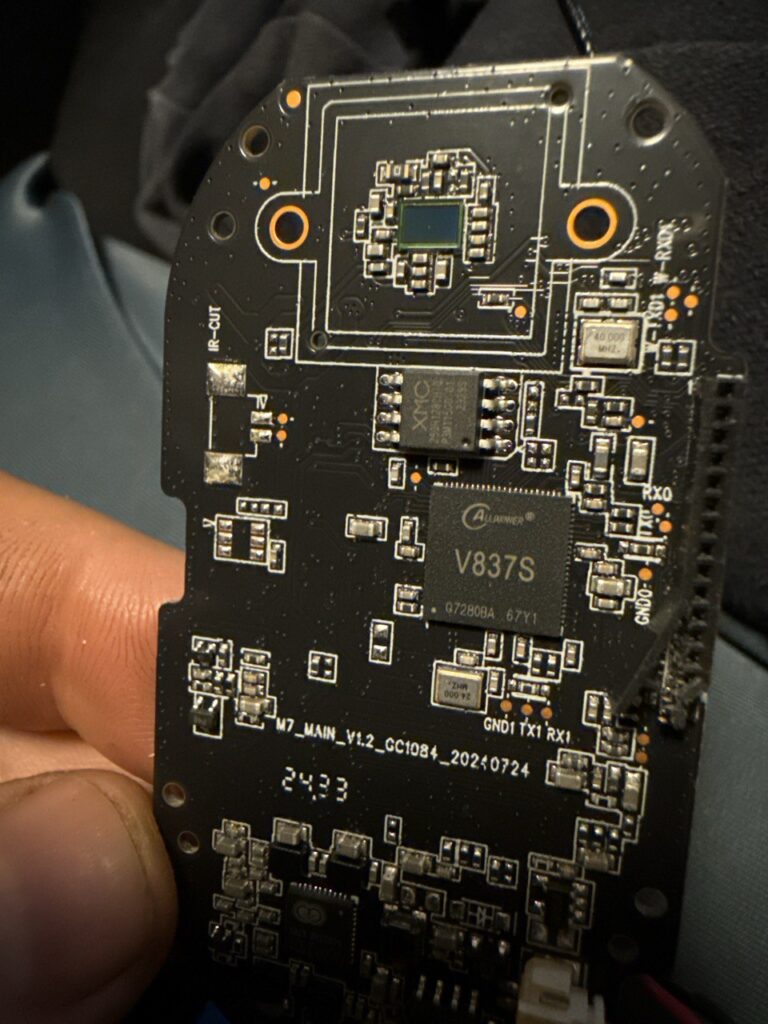
More notes.
The pins on the top right of the board above labeled W-RX0 etc. show an interface which responds only during boot, here is some sample output:
[74648][D]PACKET_CMD_USER_DATA 5a 51 0 0 64 d2 66 24
[74649][D]PACKET_CMD_USER_DATA 5a 51 0 0 64 d2 66 24
[74724][D]ecd.poweron_qrcode 0, ecd.ring_power_off_valid_count 0
[75051][I]GET_WIFI_DATA(3) len: 9 1
[75052][I]GET_NET_INFO(25) len: 8 0
$>Welcome to Wlan Bluetooth Console Tools ......
$>[75202][D]PACKET_CMD_USER_DATA 5a 51 0 0 64 d2 66 24
[75202][I]GET_SOME_DATA(7) len: 8 0
[75724][D]ecd.poweron_qrcode 0, ecd.ring_power_off_valid_count 0
[75724][I][W0] wifi: 0 81 0 | 335 137, 0(0/0/0), 0(0/0/0) | 403 0 135 112002 1042001100 | 127320 67068 60252
[UMAC WARN] sta_add():360, scan entry >= 10, discard rssi lowest one
[UMAC WARN] sta_add():360, scan entry >= 10, discard rssi lowest one
[UMAC WARN] sta_add():360, scan entry >= 10, discard rssi lowest one
[76715][D]PACKET_CMD_USER_DATA 5a 51 0 0 64 d2 66 24
[76715][I]GET_WIFI_DATA(3) len: 9 1
[76724][D]ecd.poweron_qrcode 0, ecd.ring_power_off_valid_count 0
[76724][I][W0] wifi: 0 81 0 | 169 33, 0(0/0/0), 0(0/0/0) | 403 0 135 112002 1042001100 | 127320 67716 59604
[76733][D]PACKET_CMD_USER_DATA 5a 51 0 0 64 d2 66 24
[76738][I]GET_SOME_DATA(7) len: 8 0
[net INF] msg <wlan scan success>
[76888][D]NET_CTRL_MSG_WLAN_SCAN_SUCCESS
en1: Trying to associate with 94:83:c4:6c:98:2c (SSID='youanji1' freq=2412 MHz)
en1: Associated with 94:83:c4:6c:98:2c
en1: Clear keys to send no encrypt 4 of 4-way handshake
en1: WPA: Key negotiation completed with 94:83:c4:6c:98:2c [PTK=CCMP GTK=CCMP]
en1: CTRL-EVENT-CONNECTED - Connection to 94:83:c4:6c:98:2c completed [id=0 id_str=]
[net INF] msg <wlan connected>
[net INF] netif is link up
[net INF] start DHCP...
[77267][D]============= dhcp =============
[77269][D]ip:0.0.0.0
[77271][D]netmask: 0.0.0.0
[77274][D]gateway: 0.0.0.0
[77276][D]dns[0]: 0.0.0.0
[77279][D]dns[1]: 0.0.0.0
[77281][D]dns[2]: 0.0.0.0
[77283][D]dns[3]: 0.0.0.0
[77286][D]invalid ip
[77288][D]NET_CTRL_MSG_WLAN_CONNECTED
[77724][D]ecd.poweron_qrcode 0, ecd.ring_power_off_valid_count 0
[77724][I][W0] wifi: 0 81 0 | 182 48, 0(0/0/0), 98(1/0/0) | 403 0 135 112002 1042001100 | 127320 71412 55908
[77746][D]PACKET_CMD_USER_DATA 5a 51 0 0 64 d2 66 24
[77746][I]GET_WIFI_DATA(3) len: 9 1
[77761][D]PACKET_CMD_USER_DATA 5a 51 0 0 64 d2 66 24
[77761][I]GET_SOME_DATA(7) len: 8 0
[net INF] netif (IPv4) is up
[net INF] address: 192.168.8.207
[net INF] gateway: 192.168.8.1
[net INF] netmask: 255.255.255.0
[net INF] msg <network up>
[78221][D]============= dhcp =============
[78224][D]ip:192.168.8.207
[78227][D]netmask: 255.255.255.0

 RSS
RSS