Beckman DU530 Photospectrometer notes

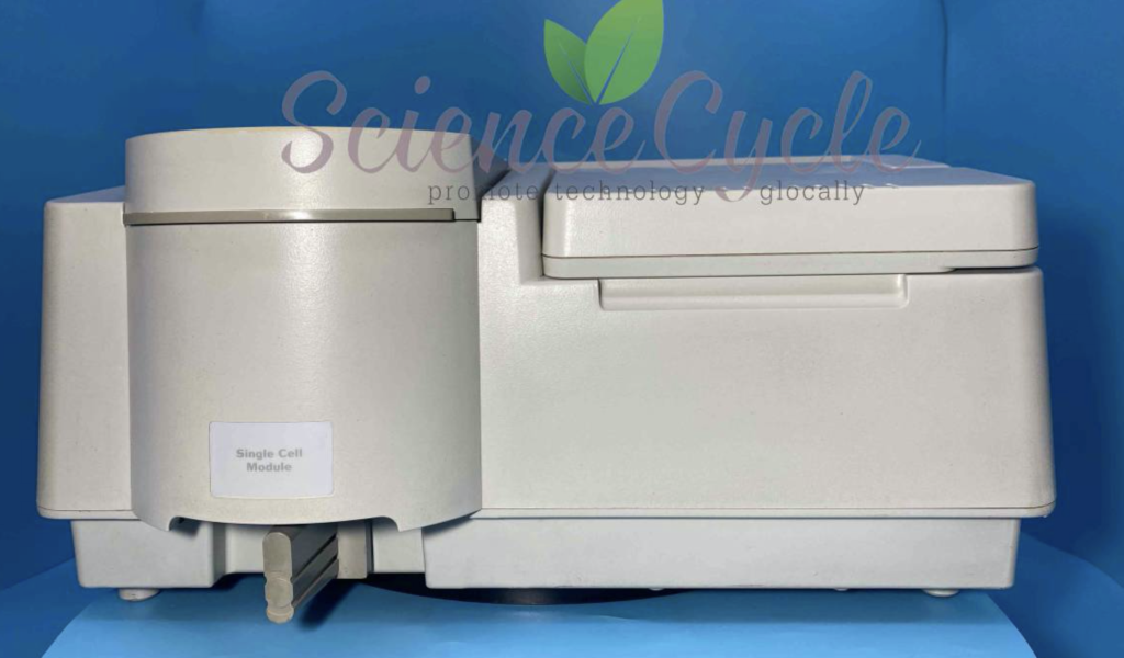
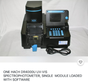
This post contains various notes on the DU530 Photospectrometer, it’s incomplete as my unit is not functioning, but never the less, my notes are here for future reference and in case they are of some use. As far as I can tell the DU 500 series is a rebadged Hach DR4000U, many of the PCBs are marked “Hach” which further supports this. And the Hach instrument looks very similar, though in stylish black:

The Hach DR4000 appears be very similar to the DU 500 Series

This is helpful, as while I’ve not been able to find a 500 Series manual online, there however a manual for the DR4000:

The DU 530 contains two light sources a small visible light lamp and a Deuterium UV lamp. This is model L2D2 and uses a 3 pin connector. The instrument appears to supply this with 12V. On my unit the Deuterium lamp appears to have failed. The unit will Pass all tests except the wavelength calibration. The unit will pass all other tests even without the lamp connected. The failure code on my unit is “24: Monochromator”.

As far as I can tell, the power on calibration doesn’t appear to try and power up the Deuterium lamp, which seems odd. It could be briefly powering the bulb and detecting a short/no current draw perhaps. It does seem to ramp up to ~600mV a few times so prehaps it’s checking the lamp resistance first. However when using the diagnostic mode (below) to fire up the lamp, it just goes straight to 12v…

It’s possible to enter a diagnostic mode when tests fail. You will be prompted for a “Service code”, after much experimentation this appears to be 3210, at least this is the case on my unit. This will let you enter a “System Check” menu. In this menu you can select various tests, for example to test motors, the keypad and display. By pressing the “SETUP” key on the keypad you can enter another menu. In this menu you can fire up the Deuterium lamp.

Overall the instrument hardware seems pretty serviceable. Only three Phillips screws hold the cover on, the keypad and some electronics are attached to the case, but cables are long enough to allow operation with the cover removed and placed to one side.

The instrument will pass all tests up to Lamp Alignment without any lamps installed. Lamp alignment will pass using only the visible lamp (and I suspect if there’s no issue all tests will pass using the visible lamp only). The instrument doesn’t seem to have anything to detect if the lamps are disconnected (other than the fact that wavelength/alignment tests will fail).

Unfortunately, I’ve not been able to find a user or service manual for the instrument. Though manuals for similar instruments are below (600 and 700 series).

This post has some information on error code 24: https://www.labwrench.com/thread/160951/refused-calibration

“Typically,  an error code 24 required a reinitialization of the spectrophotometer using a special software program and cell holder module. This can only be done by a service engineer that actually has the software and cell holder. Before going that route you might check the condition of your visible lamp and how clean your optics are. If your visible lamp’s envelope is darkened and or bubbled, replace it and try again.”

An open source program to read serial output is available here: https://github.com/Schallaven/Beckman-DU-520-reader (I have not tested this). With the tests failing, I’ve not been able to see any serial output from the unit during boot. I wonder if there’s some way to enable a serial debug interface…

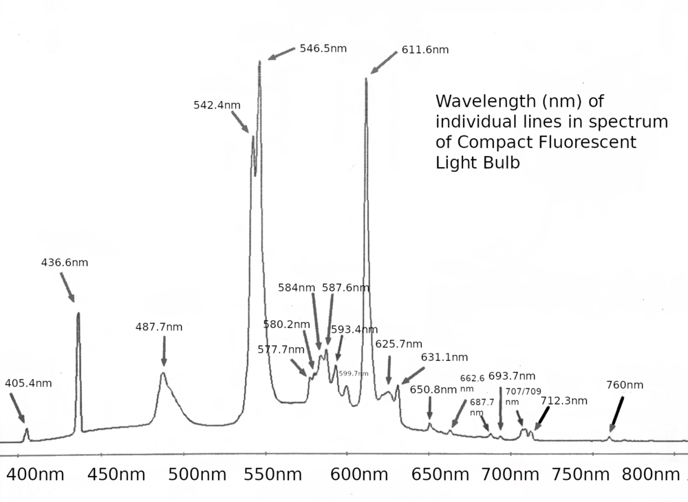
Datasheet for UV lamp used in this instrument:

DU 700 Series Manual: https://physiology.case.edu/media/eq_manuals/eq_manual_beckman_du700_.pdf

DU 600 Serial Manual: https://www.dbi.udel.edu/wp-content/uploads/2017/07/beckman-du-600-series-.pdf

ADC Datasheet: https://www.mouser.com/datasheet/2/76/cirrs01119_1-2263004.pdf

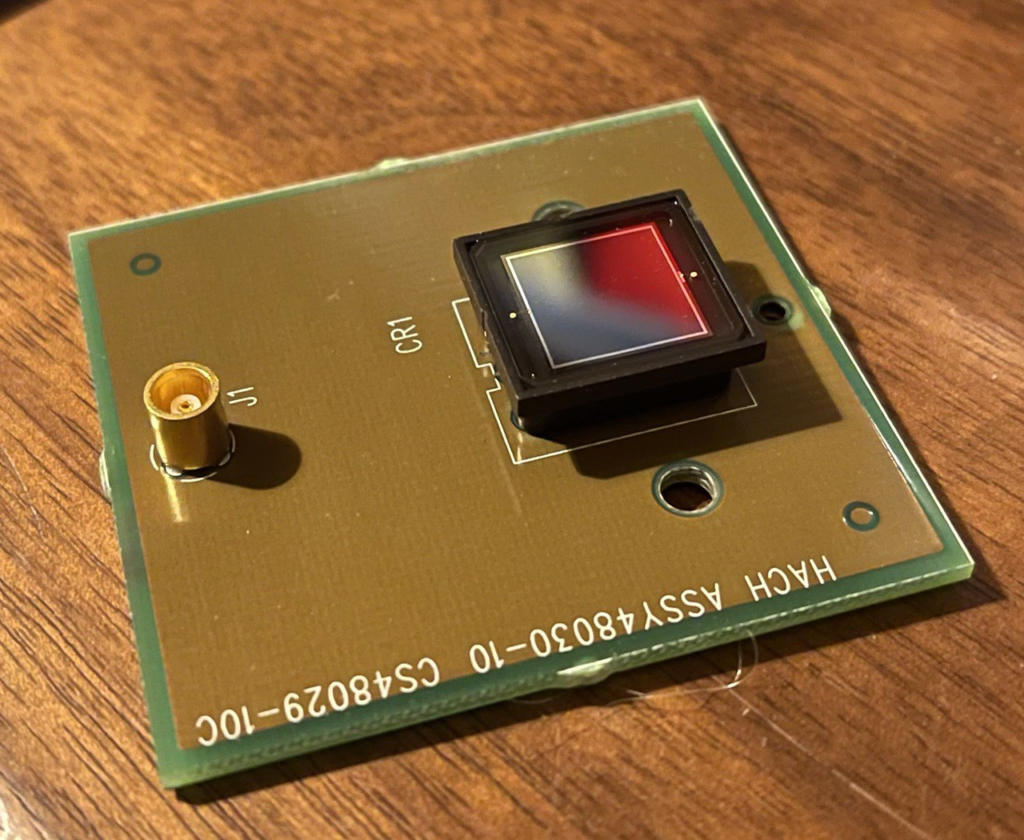
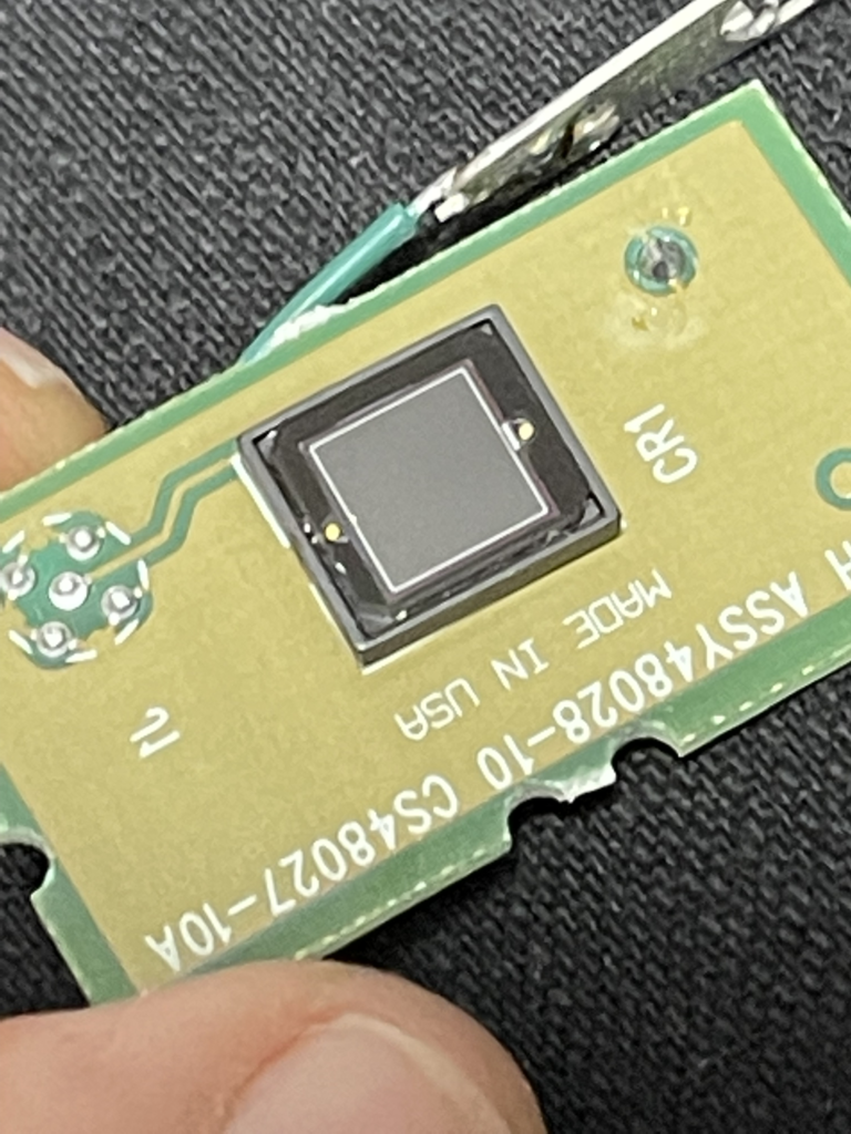
Photodiode (output side): Best I can tell is this is a S227-1010BQ, which specs sensitivity down to 190nm. Datasheet here: https://www.hamamatsu.com/resources/pdf/ssd/s1227_series_kspd1036e.pdf

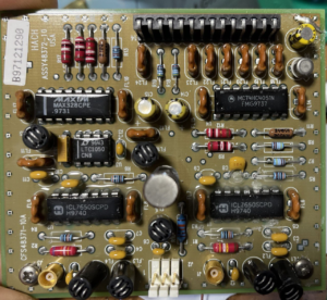
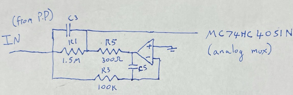
Further notes on the photodiode amplifier circuit are here.

Tested the photodiodes with a transimpedance amplifier, un-biased:

Under ambient light the small (input side) photodiode outputs about 0uA and >450uA under illumination.

Under ambient light the large (output side) photodiode outputs about 20uA and > 450uA under illumination.

Steppers used on the lamp alignment mirror and grating are Oriental Motor, Vexta PX243M-03AA-C6 and PX243M-03AA-C9, 2-phase 0.9deg/step. DC 12V 0.3A. Datasheet: https://www.orientalmotor.com/products/pdfs/opmanuals/HM-601-17JECK.pdf

Pics of various parts of the instrument below:

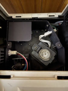
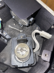
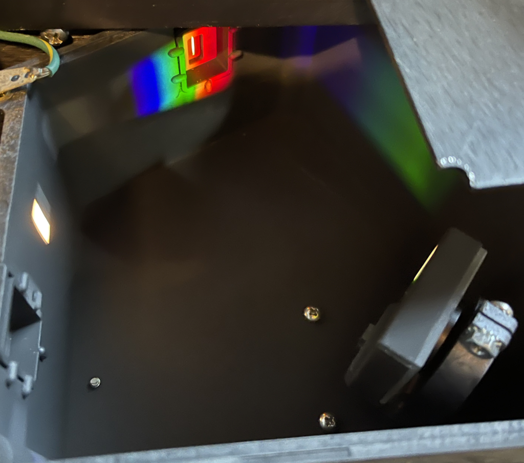
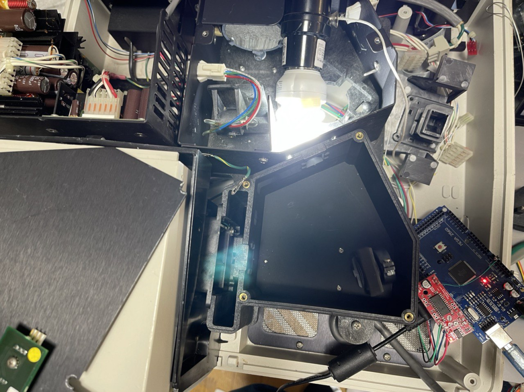
Lamp box, easily accessed without disassembly. The large lamp is the Deuterium lamp. The visible lamp is to the bottom left.
Here you can see the lamp alignment mirror (which is motorized). I assume this combines the emissions from both lamps.
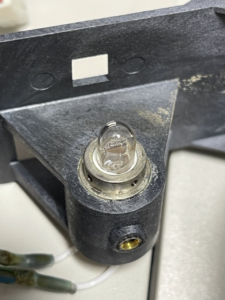
The smaller visible light lamp
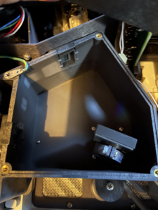
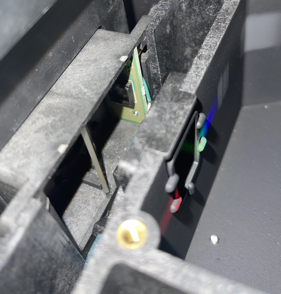
This is the monochromater block, the diffraction grating is to the lower right. The top cover of this block has what I believe is a Hall effect sensor attached. I assume this is for determining the filter wheel position (though it seems relatively far from the filter wheel).
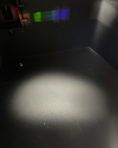
Pretty rainbows
There seems to be a beam splitter and photodiode on the output of the monochromator, there’s a filter wheel here too.
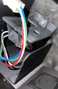
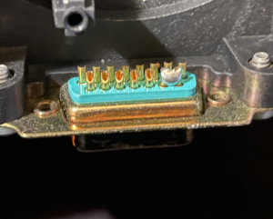
There’s a connector on the cell module, which the instrument uses to detect it’s presence (and I assume type), just a short between a couple of pins…
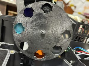
Filter wheel, many of the filters are cloudy which I suspect is the main problem with this instrument…
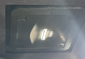
Lamp image of the slit post lamp alignment. I assume it’s essentially projecting the filament image on the slit?
Visible light lamp, it seems to be glued into the carrier.
Photodiode from the “output” side of the cuvette. Best I can tell is this is a S1227-1010BQ: https://www.hamamatsu.com/resources/pdf/ssd/s1227_series_kspd1036e.pdf
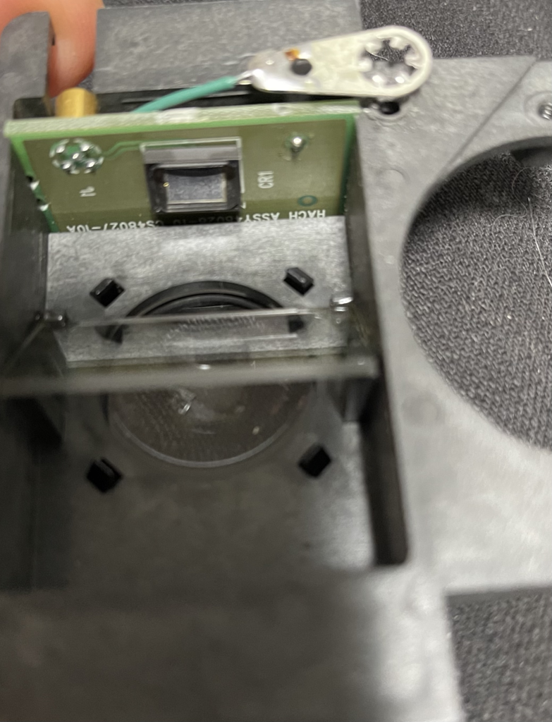
The smaller (cuvette input side) photodiode and beam splitter.
The input side photodiode
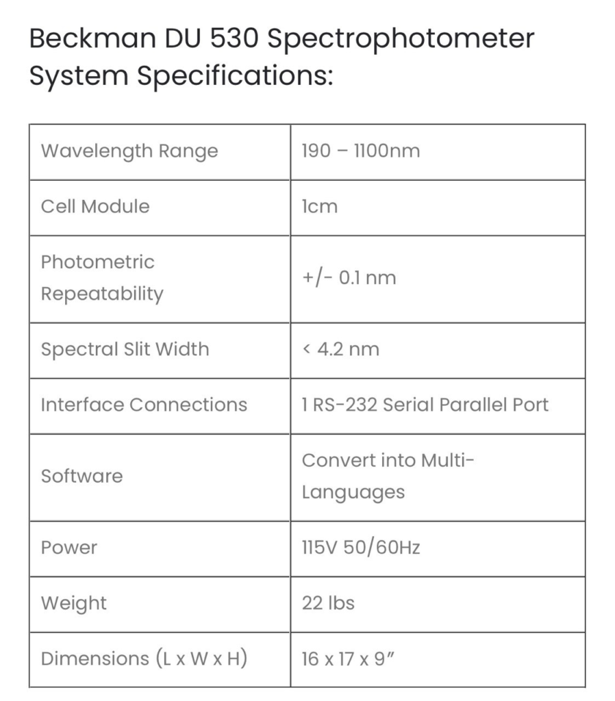
Specs found online
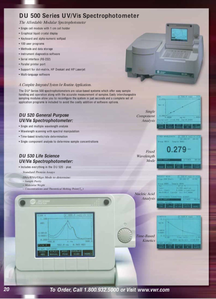
Marketing Info

Nooma Bio

This post previously appeared on my substack.

I was recently asked about Nooma Bio. This is a company and approach I’ve not looked at for some time. But it appears they published a couple of papers at the end of 2019. So here I’ll review the company and their 2019 publication.

I recently wrote about Solexa and much like that company, Nooma’s journey has taken a somewhat meandering coarse. The company was originally founded as TwoPoreGuys by Daniel Heller and William Dunbar in 2011. Dan came from a background in tech, having previously founded email software company Z-code which was acquired for ~9.4MUSD in cash and stock back in 1994.

Dan left TwoPoreGuys in 2018, and in 2019 the company turned into Ontera. Murielle Thinard-McLane took over as CEO. Nooma.bio was then founded in 2020, as a spinout of Ontera, and retains the same CEO and CTO.

Both companies are pursuing ionic solid state nanopores detection platforms. But from what I can gather, it’s Nooma that’s taking forward the original two pore approach. And this is what I’ll be discussing here.

Two Pores are better than One?

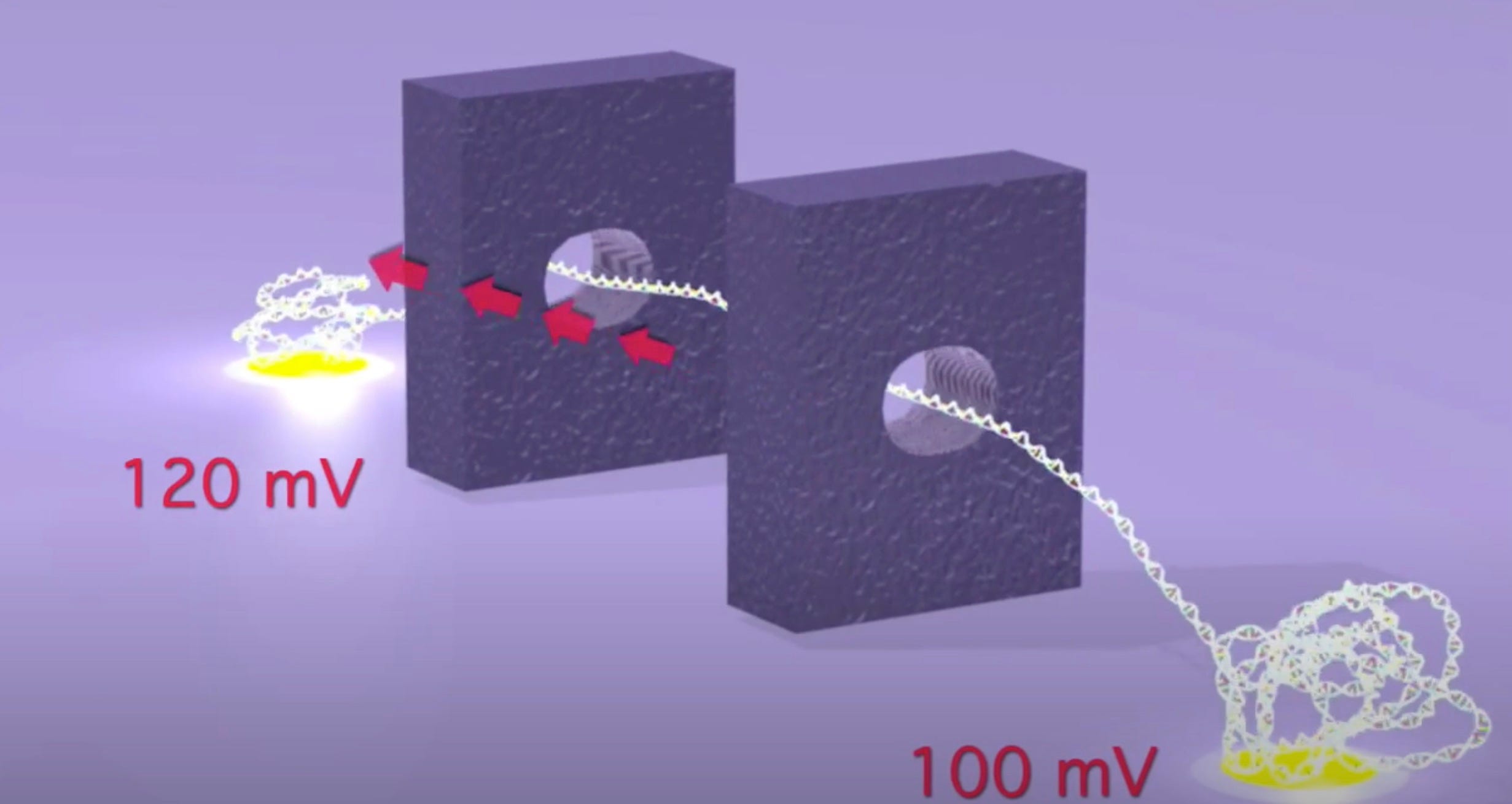
As originally presented the TwoPoreGuys’ approach used two nanopores and a small difference between two larger bias voltages across these pores. The original website, is unfortunately long gone, but the youtube videos are still up:

As presented, this never made much sense to me. The above seems electrically the same as setting a 20mV bias voltage. The two pores in this original approach were also purely for motion control, the idea was that there would be other sensors (they give the example of tunneling current electrodes) in the gap between the pores.

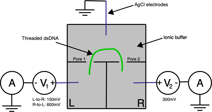
Ontera’s (pre-Nooma) 2019 paper takes basic approach in a slightly different direction. Here they use two adjacent pores on a planar substrate, in a three electrode system:

A double stranded (negatively charged) DNA translocates toward the most positive electrode. By changing the bias voltage at V₁ we can the most positive point either V₁ or V₂. All our ionic currents on the other hand will flow between V₁ / V₂ and our ground point which is in the chamber between the two pores.

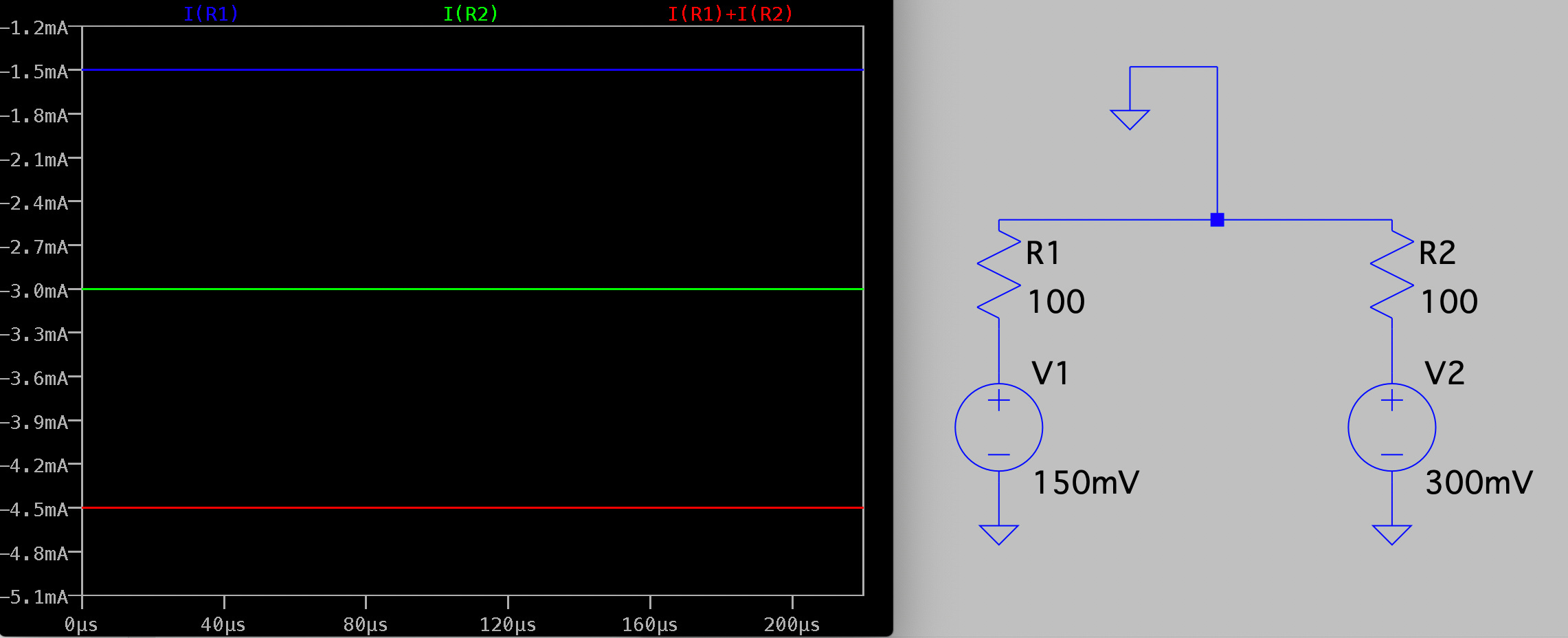
Electrically the system is pretty simple. In the diagram below R1 and R2 represent our two pores, and in reality are in the Teraohm range:

This approach gives us three principal advantages over a single pore system:

  1. We can sense the DNA twice (once as it passes through each pore).
  2. We can flip the voltages around to reverse the DNA translocation.
  3. We might get better motion control by confining the DNA between two pores.

In particular the ability to “floss” a single strand backward and forward through the pores gives multiple observations of the same molecule. You can of course also do this with single pore systems, but the author’s suggest that the two pore approach helps maintain the strands orientation.

To demonstrate this in their 2019 paper they bind a couple of Streptavidin protein tags to Lambda DNA. As these tags pass through the pore they show sharp dips in current as they block the pore. As soon as two tags have been detected, the voltage is flipped and the DNA will translocate in the back through the pores, in the reverse direction.

They can floss the same strand hundreds of times. However, it seems like there’s a fixed probability a tag will be miss registered, and the strand being ejected from the pore. This results in an exponential distribution of events/per strand (37% of the events had less than 5 scans):

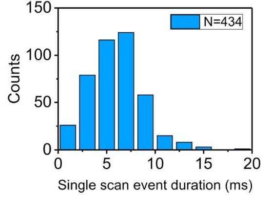
While the individual scan duration (which I think is a rough proxy for tag-to-tag dwell time), looks roughly poisson:

The paper shows a number of different experiments, with up to 7 tags at nick sites on Lambda DNA. 

Thoughts

The approach described might be usable for mapping applications. By averaging the multiple “flossed” observations you can get better estimates of the tag to tag distance. The resolution here seems like it’s on the order of a few hundred nucleotides.

So, you can imagine a platform where you nick and tag DNA and read it on a 2 pore platform. The problem is, that we already have pretty reasonable mapping tools. And the market (~$10M?) may not justify the development costs.

Unfortunatley, to me the approach doesn’t seem to be compatible with DNA. The method doesn’t slow translocation sufficiently to be able to detect single bases. In the paper, they use a 10KHz bandwidth, and Lambda strands seem to translocate in ~10ms. Which is less than 1 data point per base.

And if DNA is problematic, using this as a nanopore protein sequencing platform is likely even more challenging.

Increasing the bandwidth much beyond 10KHz isn’t very practical, and it’s not clear that you can slow the translocation (particularly of single stranded DNA) much more using this approach.

In any case, solid state pores have not yet reached feature sizes where DNA sequencing becomes practical, and this 2pore approach doesn’t seem like it would be compatible with protein nanopores and enzymatic motion control.

I do like the fact that you’re precisely stretch the strand between two points, and that you can obtain information on the strand from a first pore, before it translocates through a second. There’s one patent from Nooma discussing Material Sorting Applications which seems like an interesting idea, that could take advantage of this unique feature.

In any case, I’ll be keeping an eye on Nooma. It seems like they’ve developed an attractive technique in search of a compelling application.
Nautilus Prospectus Review

This post originally appeared on my substack.

My previous post on Nautilus covered their published IP and was originally released to paid subscribers. After it was released publicly, someone forwarded me the Nautilus’ prospectus. While the information in the prospectus is largely in line with that presented previously, it provides more concrete information on their implementation.

In the prospectus they describe “scaffolds” these are likely the SNAPs (nanoballs) from their patents. The scaffold prep process sounds non-trivial as they state flowcell/sample prep takes ~2 hours.

They also make it clear that their current chips have 10 billion sites. This is interesting because it helps frame the value of their arraying approach. To put this in context the Hiseq 2500 was able to reach 4B reads without a patterned flowcells/arraying. So, while the arraying technology is interesting it still doesn’t seem of fundamental importance to getting the platform working. The arraying approach seems to work well though, with >95% of sites being active:

There are a couple of statements on cycle requirements in the document that appear slightly contradictory. The first is “A typical 300 cycle run will generate approximately 20 terabytes of data” the second is “it takes roughly 15 cycles of multi-affinity probe biding events to uniquely identify a protein”. If you only need 15 cycles, why do you typically run to 300?

They present this plot showing classification of a protein using multi-affinity reagents:

My guess is that there are multiple sets of multi-affinity reagents. So it’s more like out of this set of 300 reagents we can find 15 that will give a unique signal for a particular protein. If this is true, then the 15 cycles statement isn’t very meaningful. And it sounds like you need to do 300 cycles in general. This implies a complex reagent cartridge and fluidics system. They state that “Nautilus intends to utilize over 300 complex reagents and various antibodies” which backs this up.

But it seems like proteins stay attached though these long runs (<1% loss):

Beyond this, they don’t say anything regarding affinity reagents other than they can use a “wide variety of “off-the-shelf” affinity reagents” and “we have developed a proprietary process for high throughput generation and characterization of multi-affinity probes”. I’d guess the high throughput generation approach is likely the sequencing based aptamer evolution approach described in their patents.

On the commercial side, they appear to be targeting a 2023 launch. So it will likely take some time for us to find out how well the platform works in practice. 

Pricing is less clear but they say it will be “in-line with mass spectrometry system budgets allocated for broad scale proteomics applications, and thus with a premium instrumentation average selling price”. However I suspect the consumables pricing will look pretty different to mass spec. That 300 reagent cartridge and patterned flowcell doesn’t sound like it’s going to be cheap.

QuantumSi Prospectus Review

This post originally appeared on the substack.

previously looked at QuantumSi’s protein sequencing approach back in September. But recently someone forwarded me their prospectus. Having recently reviewed Nautilus it seems like a good idea of revisit QuantumSi. In this post I provide an update based on my previous thoughts but you may want to refer to that post for details from their patents.

Technology

The basic process QuantumSi use to sequence proteins can be briefly described as follows:

  1. Fragment proteins into short peptides, and isolate in wells.
  2. Attach a label to the terminal amino acid, and detect the label.
  3. Remove a single terminal amino acid.
  4. Go to step 2 to identify the next amino acid.

At a high level is not unlike single molecule sequencing-by-synthesis, in that monomers are detected sequentially. The difference here being that rather than incorporating monomers, in this approach they are cleaved. QuantumSi appear to fragment the proteins prior to sequencing. I assume this is to avoid secondary structure issues. But it does mean they are getting fragmented sequences rather than an end-to-end sequence for the entire protein.

When I reviewed their patents, it was reasonably clear that you’d be unlikely to get an accurate protein sequence. It’s more likely to be a fingerprint. This means that rather than being able to call a “Y”, you’d likely be able to say this amino acid is one of “Y,W or F”.

The Prospectus suggests that can resolve these ambiguities by looking at transient binding characteristics. The “affinity reagents” they use don’t bind and stay attached. Rather they have on/off binding. So you’ll see them attach, generate a signal, then detach, then another one bind etc. Ideally a reagent that binds to “Y,W or F” might bind more strongly to one (e.g. Y) than another (e.g. W) and you can use that information to infer the amino acid type.

As mentioned in my previous post, they use fluorescent lifetime determine which affinity reagent is bound. So for every detection event they have two pieces of information, the affinity reagent type (from fluorescence lifetime, and intensity) and the binding kinetics (from the on/off rate). They call this 3 dimensional data (fluorescence life time, intensity, and kinetics).

The nice thing about this is that while you will need various reagent types, you don’t need a complex fluidic system and you are observing, and classifying them in real time. 

However, I’ve not seen anything that suggests the classification works well enough to give the full sequence. And they state that this “will ultimately enable us to cover all 20 amino acids”. Suggesting that they currently can’t.

Overall, the above approach is in line with my previous speculation based on their patents.

Chips

Like Ion Torrent, they make a big deal out of using semiconductor fabrication for their sensor: “similar to the camera in a mobile phone, our chip is produced in standard semiconductor foundries”. I generally take issue with this argument. Semiconductor fabrication is great. But if you can’t reuse the sensors it’s more like buying an expensive camera, taking one picture, then throwing the camera in the trash.

This isn’t to say that semiconductor sensing isn’t interesting… but there are other issues that need to be considered. They also talk about Moore’s law, suggesting that if “Moore’s Law remains accurate, we believe that single molecule proteomics…will allow our technology to run massively parallel measurements”. Aside from Moore’s law clearly being in trouble, this doesn’t make much sense, as there are other physical limits involved here.

From various public images, I’d guess the chip is ~15 to 20mm². PacBio chips (which use a very similar approach) have ~8M wells on a chip that appears to be roughly the same size. I didn’t see an explicit statement on read count, other than “parallel sequencing across millions of independent chambers”. But my best guess would be in the 10M range.

This puts them at the low end of throughput as compared with Nautilus and other next-gen proteomics approaches.

Product

The product has 3 components. A sample prep box (Carbon) the sequencing instrument (Platinum) and a Cloud based analysis service. Unlike Nautilus they suggest that primary data analysis happens on instrument. The instruments combined pricing is supposed to be in the $50,000 range, which is relatively cheap.

Commercial Stuff

QuantumSi say they have already initiated their early access program, but I’ve not heard of anyone else talking about this publicly. They are aiming for a commercial launch in 2022. And say that their addressable market is $21 billion. This breaks down as follows:

Of this, I think the true addressable market is closer to the $5B legacy proteomics segment. It doesn’t seem realistic to use the proposed approach for health care/diagnostics in its current form. Partly because the per-sample COGS is likely pretty high, and partly because for these applications you may want a higher throughput instrument.

They also suggest that in the future they will release lower cost instrument for at home testing:

Conclusion

QuantumSi’s approach is closer to sequencing than Nautilus, but I suspect the platform will still not give a true amino acid sequence when initially released. For the reasons highlighted in my previous post protein sequencing is just much much harder than DNA sequencing. So, like Nautilus what they’re developing may be more of a protein fingerprinting device, where traces are compared against a database of known proteins.

This begs the question: what’s the value in a relatively low throughput protein fingerprinting instrument? Where exactly the throughput spec needs to be set to be useful, particularly for diagnostic applications isn’t clear to me. But 10 million reads would certainly seem to be on the low end. I’ll try and address this in a future post.