Some Random Filters from eBay

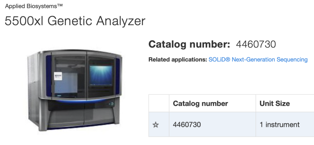
I picked up some ASI FW1000 filter wheels from eBay that were going cheap. One of them was fitted with unspecified filters. Given that a number of the FW1000s on eBay are from Genome Analyzers I suspected these would be the same as those I already have. But they were completely different.

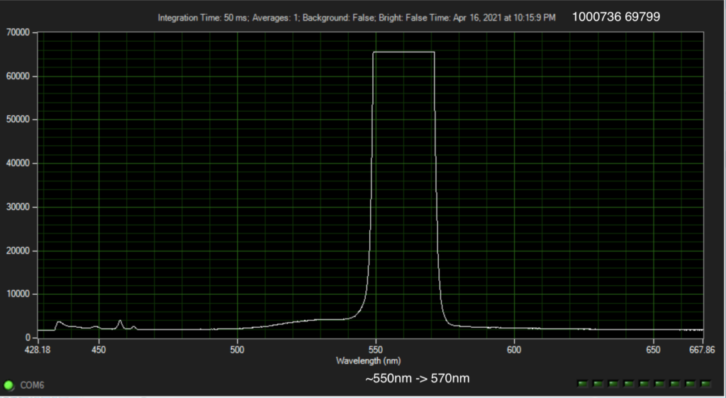
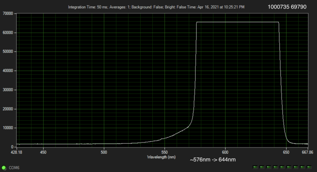
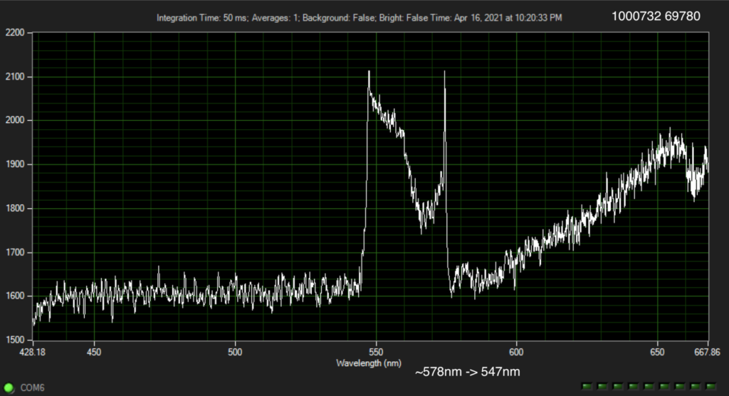
So I stuck them up against my Xenon light source and looked at the spectra… the plots are shown at the bottom of this post. Two of the filters have spectra that seem easy to interpret. 1000736 69799 (A) seems to be a band pass between ~550nm and 570nm. And 1000735 69790 (B) seems to be a band pass between ~576nm and 655nm.

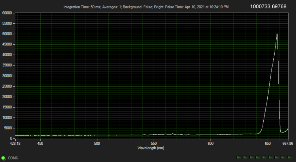
The other two, I don’t understand. 1000732 69780 is a bandpass over roughly the same range as (A) above. But seems to strongly attenuate the signal. 1000733 69768 has some kind of peak around 660nm? But it’s much less clear.

Googling around I found the following reference to filters with similar serial numbers:

These three filters match the recorded spectra well, but what exactly 1000732 is doing is less clear to me. Shame the documentation doesn’t list the manufacturer, which might yield more details…

Genome Analyzer TIRF Alignment

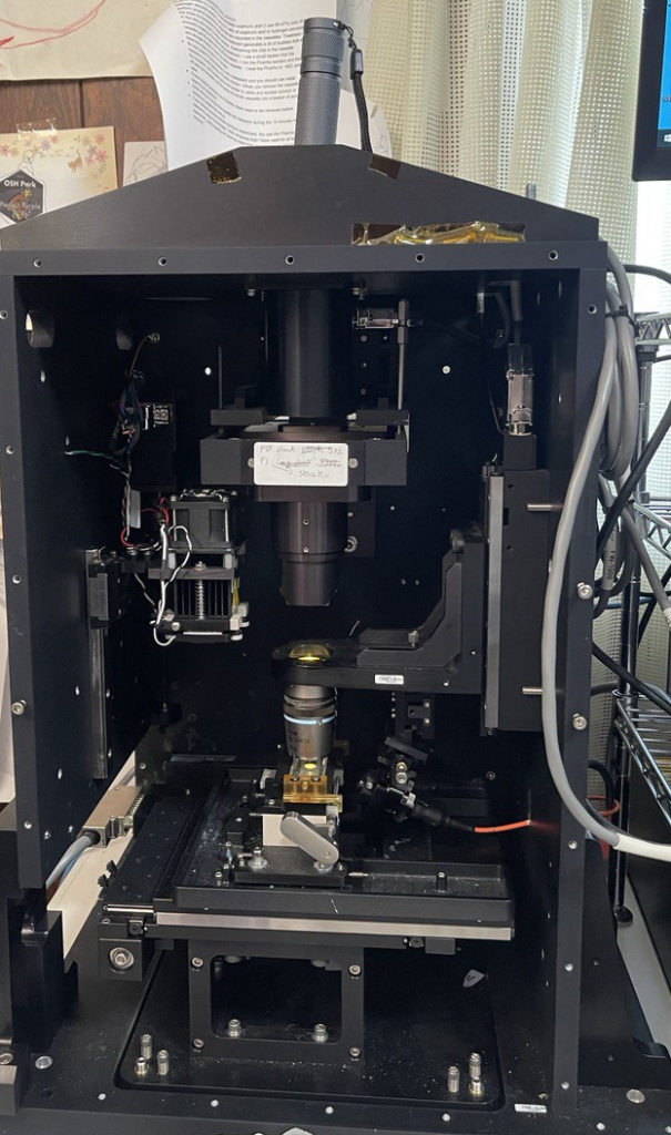
In this post I’ll document the TIRF alignment procedure I use on the Genome Analyzer 2. There doesn’t seem to be any documentation on this, so I may or may not be doing this correctly. But it’s the alignment procedure I’ve used so far…

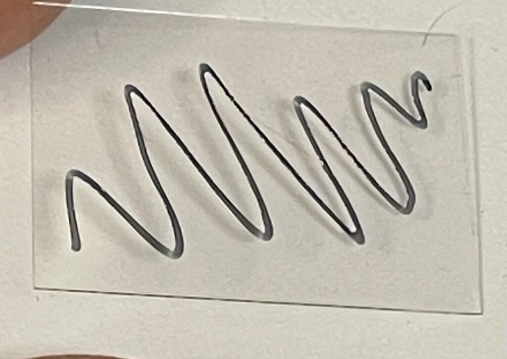
First I prep some cover glass, marking the surface, (18x24mm cover glass (Matsunami Glass Ind. Ltd.)):

I then put a drop of Cargille #19569 on the back side and place it on the prism. The prism has previously been cleaned down with Ethanol/Kimwipes:

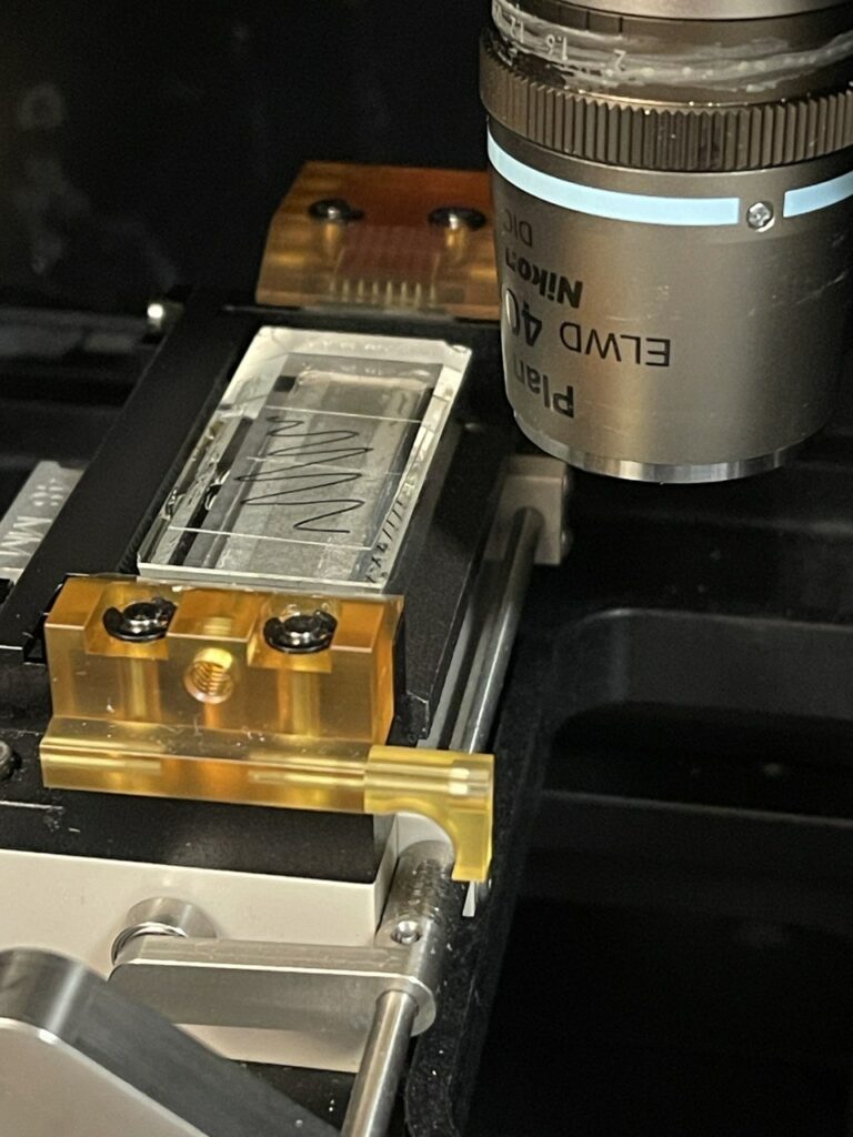
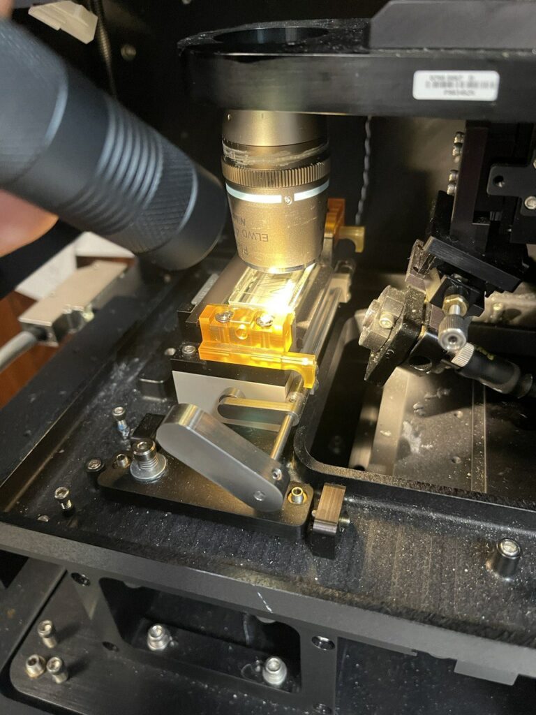
Next I move the slide under the objective and attempt to focus on the surface/marker lines, while illuminating the surface with a torch (all focusing is through micro manager/the camera, manually moving the Z-stage):

It’s important to get the objective focused on the correct plane before alignment the TIRF laser, because the TIRF laser is connected to the Z mount. As such, changes in focus will also move the laser out of alignment.

I then remove the camera and stick a torch in the C-mount hole. This shines down through the objective and you should be able to see a spot on the bottom of the prism:

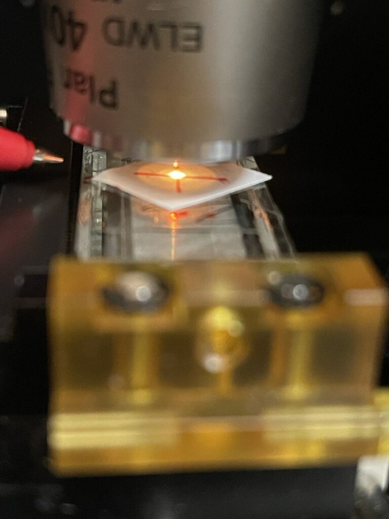
Then I draw an X on a piece of paper and place it under the objective. I align it so that the objective focal point is centered on the “X”:

You can optionally at this point, move the objective up, remove the objective and move it back down again (ensuring you move the same distance). This will let you align to the “X” with the objective out of the way…but isn’t really necessary with a long working distance objective.

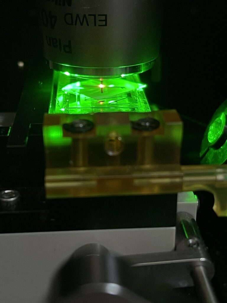
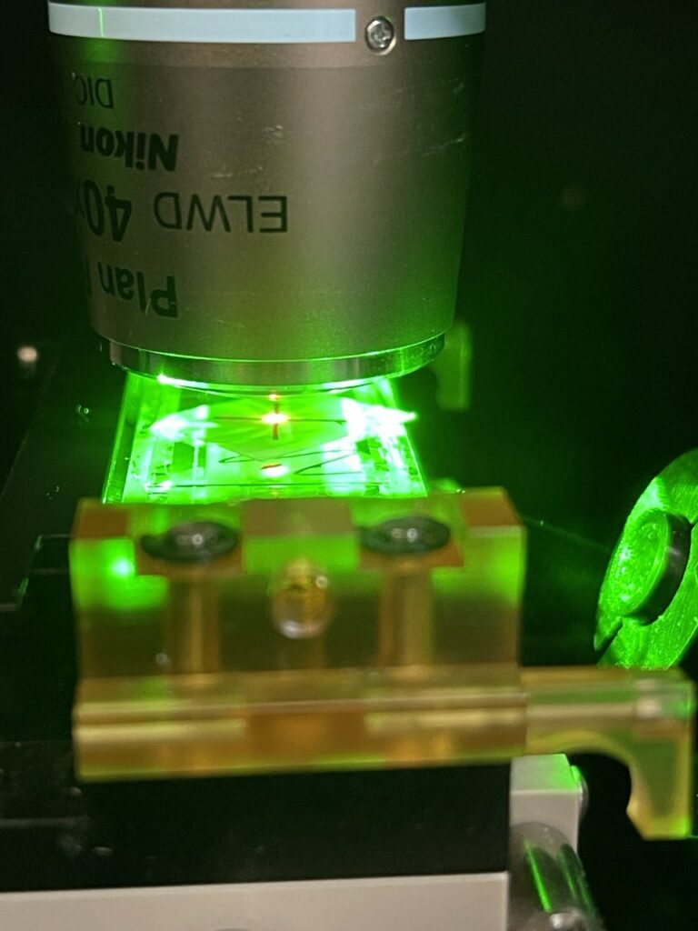
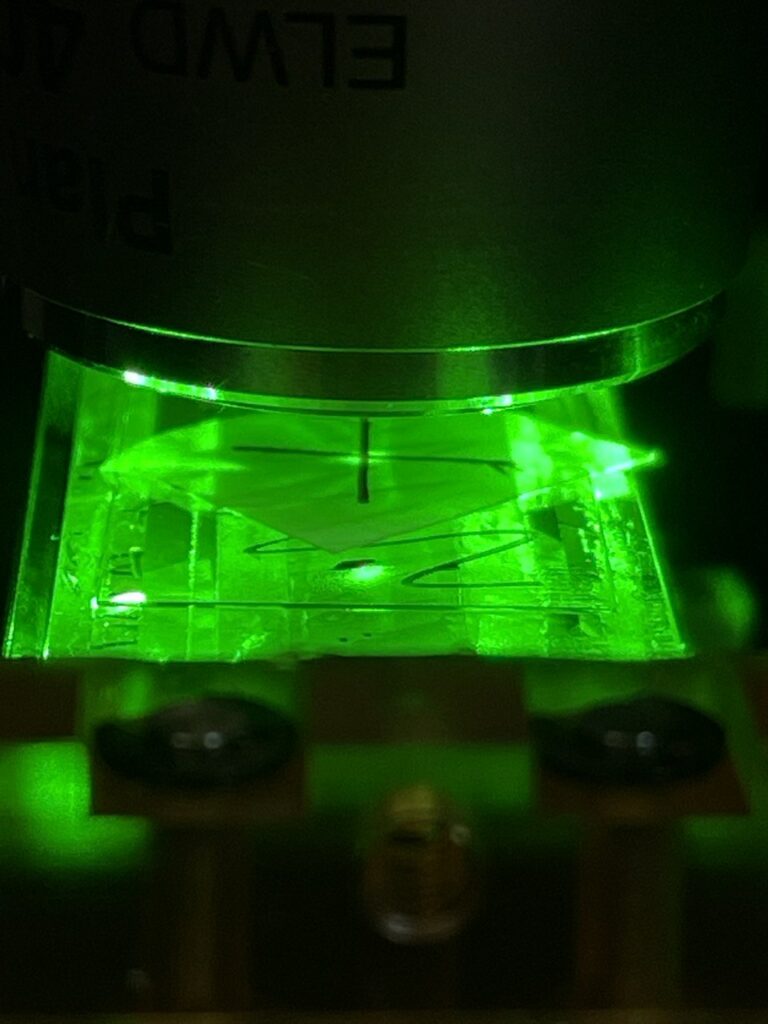
Now I turn on the TIRF laser, you can use a low power for this (say 50mW), and it makes sense to be wearing suitable eye protection. In the image below you can see the TIRF spot to the right and the objective spot in the center of the “X”:

Then, adjust the TIRF Kinematic mount until the TIRF spot is aligned with the objective spot:

You have some flexibility in terms of tuning the TIRF angle, but I’ve not experimented with this much. As a final check, it makes sense to turn of the torch (removing the objective spot) and checking that the laser is aligned with the “X”:

You can now remove the cover glass, clean the prism and everything should be aligned. The Z-axis should also be at roughly the correct focal distance, the prism will be ~170 microns down from this.

You may find that some finer adjustment is useful when performing an experiment (in my experience, it doesn’t seem to help much).

Photobeaching on the Genome Analyzer, using a IMX178

In my previous post, I talked about my first experiments with a genome analyzer, looking at single dye bleaching. In this post I discuss a few further developments.

I picked up an IMX178 based camera. These cameras are cheaper than the IMX174 based ones, have smaller pixels (bad) but better noise specs. Images generally looked a little better (the image below is atto1178_5) with used 1s exposures and 4x binning:

But I found turning the mode scrambler off produced much much cleaner images, with better defined points. However, this introduces the “wavey” illumination that you can see in the images below (500ms exposures here, atto1178_13):

Turns out the mode scrambler (which works by vibrating after all) was causing a lot of vibrational issues. While the mode scrambler is attached to a dampening mount, I found that stuffing a pile of kimwipes under the unit, cleaned up more of the vibrational noise:

The results are reasonably clean (Atto1178_21, 2s exposures, binning=4):

With some basic thresholding, I was able to quickly extract particles from this dataset in Fiji. In general for the images above I enhanced contrast (using the stack histogram) and background subtracted (default values, 50px window). Below I used analyze particles to generate particle counts on a manually thresholded stack (same thresholds were used for all images). These looks more or less as expected for a photobleaching experiment, with the dip at the beginning caused by the image becoming saturated/some spots colliding:

You can also pick out fairly clean traces across frame for spots that bleach (Plot Z-axis profile over a spot):

Datasets:

https://41j.com/blog/wp-content/uploads/2021/04/atto1178_5.tar.gz

https://41j.com/blog/wp-content/uploads/2021/04/atto1178_13.tar.gz

https://41j.com/blog/wp-content/uploads/2021/04/atto1178_21.tar.gz

Ultima Genomics

Not much is publicly available regarding Ultima genomics. The company appears to have received >3M USD in SBIR awards in 2019 and 2020 (which is by usual standards, rather a lot). Its CEO is Gilad Almogy who was previously CEO and founder of Cogenra (a solar company). Aside from this I can’t find much information about fund raising. The company appears to have been founded in late 2016. From the SBIR application, and patents it seems clear that they are focused on DNA sequencing.

Technology

I’ve taken a very brief look at a couple of Ultima’s patents. The first patent describes an imaging system for use in with a DNA sequencing platform. The substrate is on a rotating platform:

Which is somewhat reminiscent of Ling Vitae approach. Where a fluidic system was incorporated as part of a CD-like platform. I kind of like this idea, because you can potentially image a large area, with one stable axis. After an imaging pass, you end up where you started, ready to image again. In a cyclic sequencing approach, this seems attractive.

The patent also shows what looks like real image data from an ordered array:

And even some read statistics. I’d need to read this in more depth, but I suspect this is just extrapolated from spot counts:

The second patent describes a sequence-by-synthesis (SBS) sequencing chemistry. The approach appears to be somewhat similar to standard SBS, in which nucleotides are flowed in and detected in cycles. However here (unlike Illumina) reversible terminators do not appear to be used.

This is also not a traditional single-channel/unterminated SBS platform (Ion Torrent, 454), which would incorporate a single base type in each flow.

The Ultima Genomics approach presented here, works on a population of templates (so a surface amplification approach like cluster generation, or a bead based approach would need to be used).

In the dominant embodiment, it appears that Ultima Genomics would incorporate mixtures of terminated and unterminated bases. Most positions would extend normally using a wildtype nucleotide. However a smaller fraction of nucleotides would be terminated, preventing templates from extending any further. These terminated nucleotides would be labelled (likely a fluorescent label). Allowing the sequence of a cluster to be determined from the terminating sub-population.

One obvious issue with this approach, is that with each cycle, you get an accumulation of incorporated dye. You could cleave the fluorescent labels, which seems like the obvious thing to do. But the patent suggests a different approach, essentially just monitoring the increase in fluorescent intensity. An increase in a cycle means that a base was incorporated, no increase – no incorporation.

That’s fine as it goes, but eventually the build up of dye is going to make imaging problematic. You’ll either overflow the cameras range, or get increased crosstalk from scattering etc.

To avoid that one suggestion in the patent is to periodically switch dyes. That is just move to a different dye with a different emission after a few cycles. You can then switch filters, effectively cutting out fluorescence from the accumulated dye.

Overall some aspects of this approach don’t seem particularly new. Using terminators was of course the foundation of the original Sanger sequencing approach. And mixtures of nucleotide types have previously been used for sequencing in academic work. Some of this prior work may explain why the majority of claims on this patent have been cancelled.

The chemistry itself seems more complex than those currently available… so what is the advantage? I suspect the motivation is that wildtype bases likely incorporate more efficiently. This might give some improvement over Illumina-style SBS. The fact that platforms using wildtype nucleotides (Ion Torrent, 454, Sanger) have somewhat longer read lengths (on the order of 1000 nucleotides, versus 150) supports this.

But in this Ultima approach, the loss of templates through termination will eventually limit read length. It’s also worth noting that while the other platforms mentioned above have longer reads (and in some cases lower error rates) they have not proved commercially very successful. So I’d be concerned that the added complexity of the Ultima approach doesn’t provide enough benefit over existing platforms.

That said, this is only a quick review of a couple of patents. I will be watching with interest to see how things develop.